
Biopsychology
How Neurons Communicate
Learning Objectives
- Describe how neurons communicate with each other
- Explain how drugs act as agonists or antagonists for a given neurotransmitter system
Now that we have learned about the basic structures of the neuron and the role that these structures play in neuronal communication, let’s take a closer look at the signal itself—how it moves through the neuron and then jumps to the next neuron, where the process is repeated.
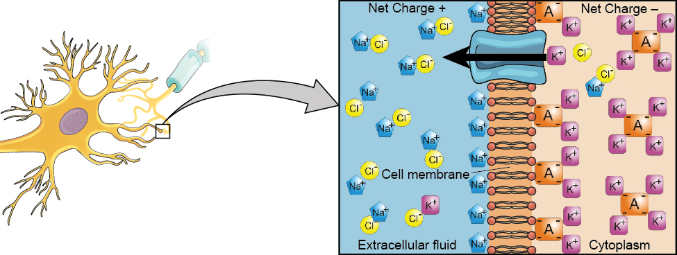
We begin at the neuronal membrane. The neuron exists in a fluid environment—it is surrounded by extracellular fluid and contains intracellular fluid (i.e., cytoplasm). The neuronal membrane keeps these two fluids separate—a critical role because the electrical signal that passes through the neuron depends on the intra- and extracellular fluids being electrically different. This difference in charge across the membrane, called the membrane potential, provides energy for the signal.
The electrical charge of the fluids is caused by charged molecules (ions) dissolved in the fluid. The semipermeable nature of the neuronal membrane somewhat restricts the movement of these charged molecules, and, as a result, some of the charged particles tend to become more concentrated either inside or outside the cell.
Between signals, the neuron membrane’s potential is held in a state of readiness, called the resting potential. Like a rubber band stretched out and waiting to spring into action, ions line up on either side of the cell membrane, ready to rush across the membrane when the neuron goes active and the membrane opens its gates (i.e., a sodium-potassium pump that allows movement of ions across the membrane). Ions in high-concentration areas are ready to move to low-concentration areas, and positive ions are ready to move to areas with a negative charge.
In the resting state, sodium (Na+) is at higher concentrations outside the cell, so it will tend to move into the cell. Potassium (K+), on the other hand, is more concentrated inside the cell, and will tend to move out of the cell (Figure 1). In addition, the inside of the cell is slightly negatively charged compared to the outside. This provides an additional force on sodium, causing it to move into the cell.
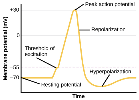
From this resting potential state, the neuron receives a signal and its state changes abruptly (Figure 2). When a neuron receives signals at the dendrites—due to neurotransmitters from an adjacent neuron binding to its receptors—small pores, or gates, open on the neuronal membrane, allowing Na+ ions, propelled by both charge and concentration differences, to move into the cell. With this influx of positive ions, the internal charge of the cell becomes more positive. If that charge reaches a certain level, called the threshold of excitation, the neuron becomes active and the action potential begins. This process of when the cell’s charge becomes positive, or less negative, is called depolarization.
Many additional pores open, causing a massive influx of Na+ ions and a huge positive spike in the membrane potential, the peak action potential. At the peak of the spike, the sodium gates close and the potassium gates open. As positively charged potassium ions leave, the cell quickly begins repolarization. At first, it hyperpolarizes, becoming slightly more negative than the resting potential, and then it levels off, returning to the resting potential.
This positive spike constitutes the action potential: the electrical signal that typically moves from the cell body down the axon to the axon terminals. The electrical signal moves down the axon like a wave; at each point, some of the sodium ions that enter the cell diffuse to the next section of the axon, raising the charge past the threshold of excitation and triggering a new influx of sodium ions. The action potential moves all the way down the axon to the terminal buttons.
Watch It
The process of neural communication is explained in the following video.
You can view the transcript for “Lights, Camera, Action Potentials!” here (opens in new window).
The action potential is an all-or-none phenomenon. In simple terms, this means that an incoming signal from another neuron is either sufficient or insufficient to reach the threshold of excitation. There is no in-between, and there is no turning off an action potential once it starts. Think of it like sending an email or a text message. You can think about sending it all you want, but the message is not sent until you hit the send button. Furthermore, once you send the message, there is no stopping it.
Because it is all or none, the action potential is recreated, or propagated, at its full strength at every point along the axon. Much like the lit fuse of a firecracker, it does not fade away as it travels down the axon. It is this all-or-none property that explains the fact that your brain perceives an injury to a distant body part like your toe as equally painful as one to your nose.
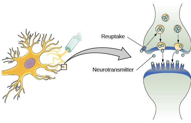
As noted earlier, when the action potential arrives at the terminal button, the synaptic vesicles release their neurotransmitters into the synapse. The neurotransmitters travel across the synapse and bind to receptors on the dendrites of the adjacent neuron, and the process repeats itself in the new neuron (assuming the signal is sufficiently strong to trigger an action potential). Once the signal is delivered, excess neurotransmitters in the synapse drift away, are broken down into inactive fragments, or are reabsorbed in a process known as reuptake. Reuptake involves the neurotransmitter being pumped back into the neuron that released it, in order to clear the synapse (Figure 3). Clearing the synapse serves both to provide a clear “on” and “off” state between signals and to regulate the production of neurotransmitter (full synaptic vesicles provide signals that no additional neurotransmitters need to be produced).
Neuronal communication is often referred to as an electrochemical event. The movement of the action potential down the length of the axon is an electrical event, and movement of the neurotransmitter across the synaptic space represents the chemical portion of the process.
Watch It
Watch the following video to see how neurons communicate within the body.
You can view the transcript for “How do nerves work? – Elliot Krane” here (opens in new window).
Neurotransmitters and Drugs
There are several different types of neurotransmitters released by different neurons, and we can speak in broad terms about the kinds of functions associated with different neurotransmitters (Table 1). Much of what psychologists know about the functions of neurotransmitters comes from research on the effects of drugs in psychological disorders. Psychologists who take a biological perspective and focus on the physiological causes of behavior assert that psychological disorders like depression and schizophrenia are associated with imbalances in one or more neurotransmitter systems. In this perspective, psychotropic medications can help improve the symptoms associated with these disorders. Psychotropic medications are drugs that treat psychiatric symptoms by restoring neurotransmitter balance.
| Neurotransmitter | Involved in | Potential Effect on Behavior |
|---|---|---|
| Acetylcholine | Muscle action, memory | Increased arousal, enhanced cognition |
| Beta-endorphin | Pain, pleasure | Decreased anxiety, decreased tension |
| Dopamine | Mood, sleep, learning | Increased pleasure, suppressed appetite |
| Gamma-aminobutyric acid (GABA) | Brain function, sleep | Decreased anxiety, decreased tension |
| Glutamate | Memory, learning | Increased learning, enhanced memory |
| Norepinephrine | Heart, intestines, alertness | Increased arousal, suppressed appetite |
| Serotonin | Mood, sleep | Modulated mood, suppressed appetite |
Psychoactive drugs can act as agonists or antagonists for a given neurotransmitter system. Agonists are chemicals that mimic a neurotransmitter at the receptor site and, thus, strengthen its effects. An antagonist, on the other hand, blocks or impedes the normal activity of a neurotransmitter at the receptor. Agonist and antagonist drugs are prescribed to correct the specific neurotransmitter imbalances underlying a person’s condition. For example, Parkinson’s disease, a progressive nervous system disorder, is associated with low levels of dopamine. Therefore dopamine agonists, which mimic the effects of dopamine by binding to dopamine receptors, are one treatment strategy.
Certain symptoms of schizophrenia are associated with overactive dopamine neurotransmission. The antipsychotics used to treat these symptoms are antagonists for dopamine—they block dopamine’s effects by binding its receptors without activating them. Thus, they prevent dopamine released by one neuron from signaling information to adjacent neurons.
In contrast to agonists and antagonists, which both operate by binding to receptor sites, reuptake inhibitors prevent unused neurotransmitters from being transported back to the neuron. This leaves more neurotransmitters in the synapse for a longer time, increasing its effects. Depression, which has been consistently linked with reduced serotonin levels, is commonly treated with selective serotonin reuptake inhibitors (SSRIs). By preventing reuptake, SSRIs strengthen the effect of serotonin, giving it more time to interact with serotonin receptors on dendrites. Common SSRIs on the market today include Prozac, Paxil, and Zoloft. The drug LSD is structurally very similar to serotonin, and it affects the same neurons and receptors as serotonin. Psychotropic drugs are not instant solutions for people suffering from psychological disorders. Often, an individual must take a drug for several weeks before seeing improvement, and many psychoactive drugs have significant negative side effects. Furthermore, individuals vary dramatically in how they respond to the drugs. To improve chances for success, it is not uncommon for people receiving pharmacotherapy to undergo psychological and/or behavioral therapies as well. Some research suggests that combining drug therapy with other forms of therapy tends to be more effective than any one treatment alone (for one such example, see March et al., 2007).
Watch It
Review the process of neural communication in the following CrashCourse psychology video:
You can view the transcript for “The Chemical Mind: Crash Course Psychology #3” here (opens in new window).
Licenses and Attributions (Click to expand)
CC licensed content, Original
- Addition of link to learning. Provided by: Lumen Learning. License: CC BY: Attribution
CC licensed content, Shared previously
- Cells of the Nervous System. Authored by: OpenStax College. Located at: https://openstax.org/books/psychology-2e/pages/3-2-cells-of-the-nervous-system. License: CC BY: Attribution. License Terms: Download for free at https://openstax.org/books/psychology-2e/pages/1-introduction.
All rights reserved content
- The Chemical Mind – Crash Course Psychology #3. Authored by: Hank Green. Provided by: CrashCourse. Located at: https://www.youtube.com/watch?v=W4N-7AlzK7s. License: Other. License Terms: Standard YouTube License
- Lights, Camera, Action Potentials!. Authored by: Carleton University. Located at: https://youtu.be/XdCrZm_JAp0?t=1m20s. License: Other. License Terms: Standard YouTube License
- How do nerves work? . Authored by: Elliot Krane. Provided by: TedEd. Located at: https://www.youtube.com/watch?v=uU_4uA6-zcE#t=64. License: Other. License Terms: Standard YouTube License
cells in the nervous system that act as interconnected information processors, which are essential for all of the tasks of the nervous system
difference in charge across the neuronal membrane
the state of readiness of a neuron membrane’s potential between signals
protein on the cell surface where neurotransmitters attach
level of charge in the membrane that causes the neuron to become active
when a cell’s charge becomes positive, or less negative
when a cell’s charge becomes more negative than its resting potential
electrical signal that moves down the neuron’s axon
axon terminal containing synaptic vesicles
phenomenon that incoming signal from another neuron is either sufficient or insufficient to reach the threshold of excitation
storage site for neurotransmitters
small gap between two neurons where communication occurs
neurotransmitter is pumped back into the neuron that released it
chemical messenger of the nervous system
view that psychological disorders like depression and schizophrenia are associated with imbalances in one or more neurotransmitter systems
drugs that treat psychiatric symptoms by restoring neurotransmitter balance
drug that mimics or strengthens the effects of a neurotransmitter
drug that blocks or impedes the normal activity of a given neurotransmitter
