
43.3 Human Reproduction and Gametogenesis
Learning Objectives
- Describe spermatogenesis and oogenesis, in mammals, and discuss their differences and similarities
As animals became more complex, specific organs and organ systems developed to support specific functions for the organism. The reproductive structures that evolved in land animals allow males and females to mate, fertilize internally, and support the growth and development of offspring.
Mammal Reproductive Anatomy
The reproductive tissues of male and female humans develop similarly in utero until a low level of the hormone testosterone is released from male gonads. Testosterone causes the undeveloped tissues to differentiate into male sexual organs. When testosterone is absent, the tissues develop into female sexual tissues. Primitive gonads become testes or ovaries.
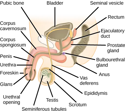
Male Reproductive Anatomy
In the male reproductive system, the scrotum houses the testicles or testes (singular: testis), including providing passage for blood vessels, nerves, and muscles related to testicular function. The testes are a pair of male reproductive organs that produce sperm and some reproductive hormones.
Visual Connection
Female Reproductive Anatomy
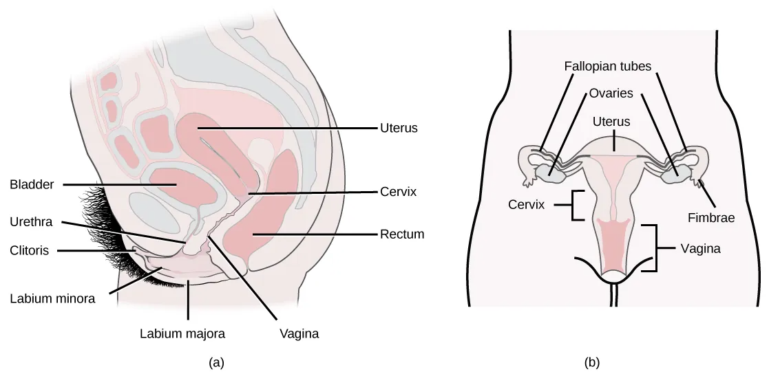
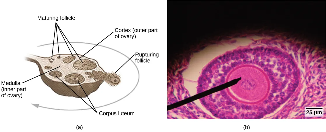
Internal female reproductive structures include ovaries, oviducts, the uterus, and the vagina, shown in Figure 43.10. Ovaries consist of a medulla and cortex: the medulla contains nerves and blood vessels to supply the cortex with nutrients and remove waste. The outer layers of cells of the cortex are the functional parts of the ovaries. The cortex is made up of follicular cells that surround eggs that develop during fetal development in utero. During the menstrual period, a batch of follicular cells develops and prepares the eggs for release. At ovulation, one follicle ruptures and one egg is released, as illustrated in Figure 43.11a.
Gametogenesis (Spermatogenesis and Oogenesis)
Gametogenesis, the production of sperm and eggs, takes place through the process of meiosis. During meiosis, two cell divisions separate the paired chromosomes in the nucleus and then separate the chromatids that were made during an earlier stage of the cell’s life cycle. Meiosis produces haploid cells with one member of each pair of chromosomes normally found in diploid cells. The production of sperm is called spermatogenesis and the production of eggs is called oogenesis.
Spermatogenesis
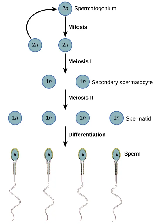
Spermatogenesis, illustrated in Figure 43.12, occurs in the wall of the seminiferous tubules (Figure 43.8), with stem cells at the periphery of the tube and the spermatozoa at the lumen of the tube. Immediately under the capsule of the tubule are diploid, undifferentiated cells. These stem cells, called spermatogonia (singular: spermatagonium), go through mitosis with one offspring going on to differentiate into a sperm cell and the other giving rise to the next generation of sperm.
Meiosis starts with a cell called a primary spermatocyte. At the end of the first meiotic division, a haploid cell is produced called a secondary spermatocyte. This cell is haploid and must go through another meiotic cell division. The cell produced at the end of meiosis is called a spermatid and when it reaches the lumen of the tubule and grows a flagellum, it is called a sperm cell. Four sperm result from each primary spermatocyte that goes through meiosis.
Stem cells are deposited during gestation and are present at birth through the beginning of adolescence, but in an inactive state. During adolescence, gonadotropic hormones from the anterior pituitary cause the activation of these cells and the production of viable sperm. This continues into old age.
Oogenesis
Oogenesis, illustrated in Figure 43.13, occurs in the outermost layers of the ovaries. As with sperm production, oogenesis starts with a germ cell, called an oogonium (plural: oogonia), but this cell undergoes mitosis to increase in number, eventually resulting in up to about one to two million cells in the embryo.
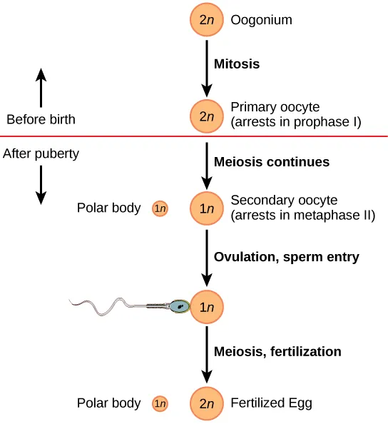
The cell starting meiosis is called a primary oocyte, as shown in Figure 43.13. This cell will start the first meiotic division and be arrested in its progress in the prophase I stage. At the time of birth, all future eggs are in the prophase stage. At adolescence, anterior pituitary hormones, follicle stimulating hormone (FSH) cause the development of a number of follicles in an ovary. This results in the primary oocyte finishing the first meiotic division. The cell divides unequally, with most of the cellular material and organelles going to one cell, called a secondary oocyte, and only one set of chromosomes and a small amount of cytoplasm going to the other cell. This second cell is called a polar body and usually dies. A secondary meiotic arrest occurs, this time at the metaphase II stage. At ovulation, this secondary oocyte will be released and travel toward the uterus through the oviduct. If the secondary oocyte is fertilized, the cell continues through the meiosis II, producing a second polar body and a fertilized egg containing all 46 chromosomes of a human being, half of them coming from the sperm.
Egg production begins before birth, is arrested during meiosis until puberty, and then individual cells continue through at each menstrual cycle. One egg is produced from each meiotic process, with the extra chromosomes and chromatids going into polar bodies that degenerate and are reabsorbed by the body.
