
24.1 Characteristics of Fungi
Learning Outcomes
- List the characteristics of fungi
- Describe the composition of the mycelium
- Describe the mode of nutrition of fungi
- Explain sexual and asexual reproduction in fungi
Although humans have used yeasts and mushrooms since prehistoric times, until recently, the biology of fungi was poorly understood. In fact, up until the mid-20th century, many scientists classified fungi as plants! Fungi, like plants, are mostly sessile and seemingly rooted in place. They possess a stem-like structure similar to plants, as well as having a root-like fungal mycelium in the soil. In addition, their mode of nutrition was poorly understood. Progress in the field of fungal biology was the result of mycology: the scientific study of fungi. Based on fossil evidence, fungi appeared in the pre-Cambrian era, about 450 million years ago. Molecular biology analysis of the fungal genome demonstrates that fungi are more closely related to animals than plants. Under some current systematic phylogenies, they continue to be a polyphyletic group of organisms that share characteristics, rather than sharing a single common ancestor.
Cell Structure and Function
Fungi are eukaryotes, and as such, have a complex cellular organization. As eukaryotes, fungal cells contain a membrane-bound nucleus. The DNA in the nucleus is wrapped around histone proteins, as is observed in other eukaryotic cells. A few types of fungi have accessory genomic structures comparable to bacterial plasmids (loops of DNA); however, the horizontal transfer of genetic information that occurs between one bacterium and another rarely occurs in fungi. Fungal cells also contain mitochondria and a complex system of internal membranes, including the endoplasmic reticulum and Golgi apparatus.
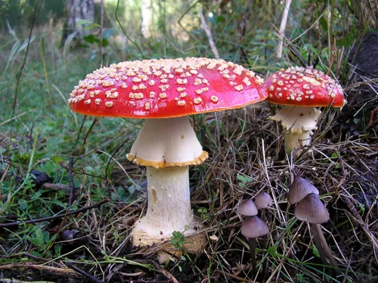
Unlike plant cells, fungal cells do not have chloroplasts or chlorophyll. Many fungi display bright colors arising from other cellular pigments, ranging from red to green to black. The poisonous Amanita muscaria (fly agaric) is recognizable by its bright red cap with white patches (Figure 24.2). Pigments in fungi are associated with the cell wall and play a protective role against ultraviolet radiation. Some fungal pigments are toxic to humans.
Like plant cells, fungal cells have a thick cell wall. The rigid layers of fungal cell walls contain complex polysaccharides called chitin and glucans. Chitin (N-acetyl-D-glucosamine), also found in the exoskeleton of arthropods such as insects, gives structural strength to the cell walls of fungi. The wall protects the cell from desiccation and some predators. Fungi have plasma membranes similar to those of other eukaryotes, except that the structure is stabilized by ergosterol: a steroid molecule that replaces the cholesterol found in animal cell membranes. Most members of the kingdom Fungi are nonmotile. However, flagella are produced by the spores and gametes in the primitive Phylum Chytridiomycota.
Growth
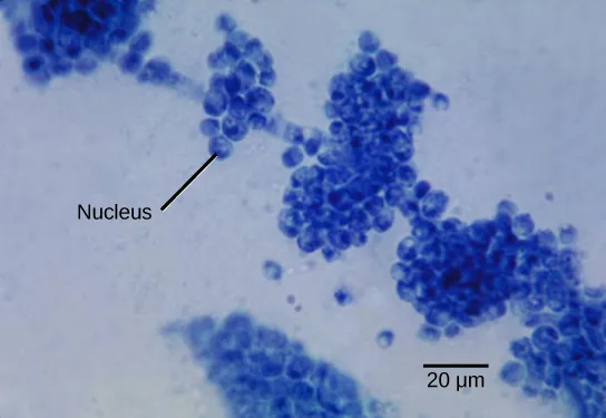
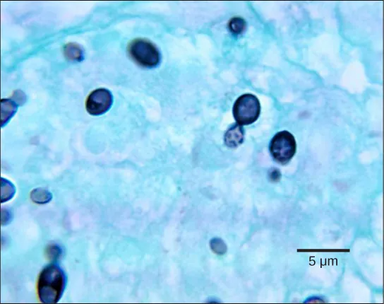
The vegetative body of a fungus is a unicellular or multicellular thallus. Unicellular fungi are called yeasts. Multicellular fungi produce threadlike hyphae (singular hypha). Dimorphic fungi can change from the unicellular to multicellular state depending on environmental conditions. Saccharomyces cerevisiae (baker’s yeast) and Candida species (the agents of thrush, a common fungal infection) are examples of unicellular fungi (Figure 24.3).
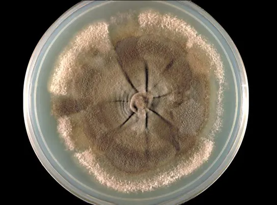
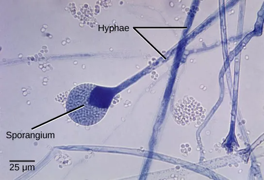
Most fungi are multicellular organisms. They display two distinct morphological stages: the vegetative and reproductive. The vegetative stage consists of a tangle of hyphae, whereas the reproductive stage can be more conspicuous. The mass of hyphae is a mycelium (Figure 24.4). It can grow on a surface, in soil or decaying material, in a liquid, or even on living tissue. Although individual hyphae must be observed under a microscope, the mycelium of a fungus can be very large, with some species truly being “the fungus humongous.” The giant Armillaria solidipes (honey mushroom) is considered the largest organism on Earth, spreading across more than 2,000 acres of underground soil in eastern Oregon; it is estimated to be at least 2,400 years old.
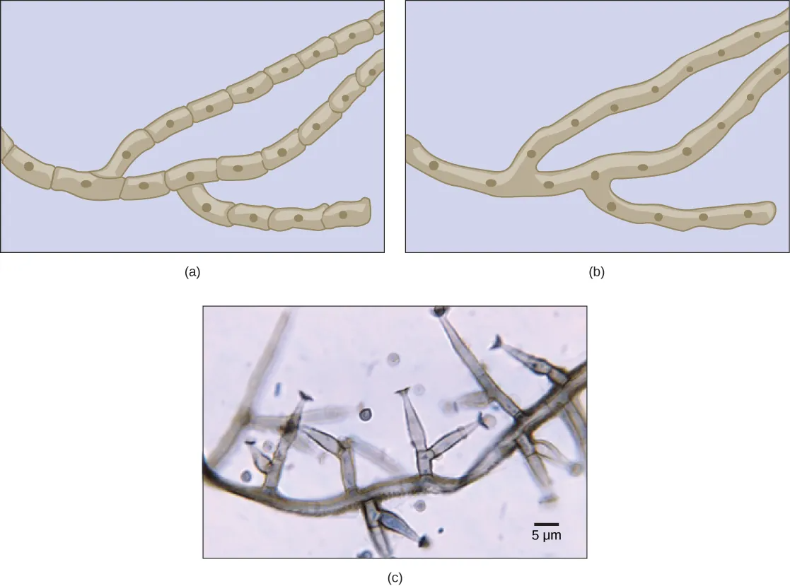
Most fungal hyphae are divided into separate cells by endwalls called septa (singular, septum) (Figure 24.5a, c). In most phyla of fungi, tiny holes in the septa allow for the rapid flow of nutrients and small molecules from cell to cell along the hypha. They are described as perforated septa. The hyphae in bread molds (which belong to the Phylum Zygomycota) are not separated by septa. Instead, they are formed by large cells containing many nuclei (multinucleate), an arrangement described as coenocytic hyphae (Figure 24.5b).
Fungi thrive in environments that are moist and slightly acidic, and can grow with or without light. They vary in their oxygen requirement. Most fungi are obligate aerobes, requiring oxygen to survive. Other species, such as members of the Chytridiomycota that reside in the rumen of cattle, are obligate anaerobes, in that they only use anaerobic respiration because oxygen will disrupt their metabolism or kill them. Yeasts are intermediate, being facultative anaerobes. This means that they grow best in the presence of oxygen using aerobic respiration, but can survive using anaerobic respiration when oxygen is not available. The alcohol produced from yeast fermentation is used in wine and beer production.
Nutrition
Like animals, fungi are heterotrophs; they use complex organic compounds as a source of carbon, rather than fix carbon dioxide from the atmosphere as do some bacteria and most plants. In addition, fungi do not fix nitrogen from the atmosphere. Like animals, they must obtain it from their diet. However, unlike most animals, which ingest food and then digest it internally in specialized organs, fungi perform these steps in the reverse order; digestion precedes ingestion. First, exoenzymes are transported out of the hyphae, where they process nutrients in the environment. Then, the smaller molecules produced by this external digestion are absorbed through the large surface area of the mycelium. As with animal cells, the polysaccharide of storage is glycogen, a branched polysaccaride, rather than amylopectin, a less densely branched polysaccharide, and amylose, a linear polysaccharide, as found in plants.
Fungi are mostly saprobes (saprophyte is an equivalent term): organisms that derive nutrients from decaying organic matter. They obtain their nutrients from dead or decomposing organic material derived mainly from plants. Fungal exoenzymes are able to break down insoluble compounds, such as the cellulose and lignin of dead wood, into readily absorbable glucose molecules. The carbon, nitrogen, and other elements are thus released into the environment. Because of their varied metabolic pathways, fungi fulfill an important ecological role and are being investigated as potential tools in bioremediation of chemically damaged ecosystems. For example, some species of fungi can be used to break down diesel oil and polycyclic aromatic hydrocarbons (PAHs). Other species take up heavy metals, such as cadmium and lead.
Some fungi are parasitic, infecting either plants or animals. Smut and Dutch elm disease affect plants, whereas athlete’s foot and candidiasis (thrush) are medically important fungal infections in humans. In environments poor in nitrogen, some fungi resort to predation of nematodes (small non-segmented roundworms). In fact, species of Arthrobotrys fungi have a number of mechanisms to trap nematodes: One mechanism involves constricting rings within the network of hyphae. The rings swell when they touch the nematode, gripping it in a tight hold. The fungus then penetrates the tissue of the worm by extending specialized hyphae called haustoria. Many parasitic fungi possess haustoria, as these structures penetrate the tissues of the host, release digestive enzymes within the host’s body, and absorb the digested nutrients.
Reproduction
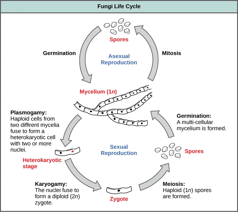
Fungi reproduce sexually and/or asexually. Perfect fungi reproduce both sexually and asexually, while the so-called imperfect fungi reproduce only asexually (by mitosis).
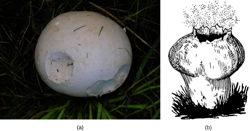
In both sexual and asexual reproduction, fungi produce spores that disperse from the parent organism by either floating on the wind or hitching a ride on an animal. Fungal spores are smaller and lighter than plant seeds. For example, the giant puffball mushroom bursts open and releases trillions of spores in a massive cloud of what looks like finely particulate dust. The huge number of spores released increases the likelihood of landing in an environment that will support growth (Figure 24.6).
Asexual Reproduction
Fungi reproduce asexually by fragmentation, budding, or producing spores. Fragments of hyphae can grow new colonies. Somatic cells in yeast form buds. During budding (an expanded type of cytokinesis), a bulge forms on the side of the cell, the nucleus divides mitotically, and the bud ultimately detaches itself from the mother cell (Figure 24.7).
Sexual Reproduction
Sexual reproduction introduces genetic variation into a population of fungi. In fungi, sexual reproduction often occurs in response to adverse environmental conditions. During sexual reproduction, two mating types are produced. When both mating types are present in the same mycelium, it is called homothallic, or self-fertile. Heterothallic mycelia require two different, but compatible, mycelia to reproduce sexually.
Although there are many variations in fungal sexual reproduction, all include the following three stages (Figure 24.8). First, during plasmogamy (literally, “marriage or union of cytoplasm”), two haploid cells fuse, leading to a dikaryotic stage where two haploid nuclei coexist in a single cell. During karyogamy (“nuclear marriage”), the haploid nuclei fuse to form a diploid zygote nucleus. Finally, meiosis takes place during which gametes of different mating types are generated. At this stage, spores are disseminated into the environment.
Link to Learning
Characteristics of fungi
Review the characteristics of fungi by visiting this interactive site from Wisconsin-online.
Link to Learning
Fungi, a recap
This video by Crash Course recaps some of the basics regarding the Kingdom of the fungi.
Table of Contents
- Biolography 02:07
- Structure 04:53
- The Decomposers 06:10
- The Mutualists 06:38
- The Predators 07:23
- The Parasites 07:35
- Reproduction 08:24
scientific study of fungi
vegetative body of a fungus
general term used to describe unicellular fungi
fungal filament composed of one or more cells
mass of fungal hyphae
cell wall division between hyphae
single hypha that lacks septa and contains many nuclei
organisms, such as humans, that must perform aerobic respiration to survive
organisms that only perform anaerobic respiration and often cannot survive in the presence of oxygen
organisms that can perform both aerobic and anaerobic respiration and can survive in oxygen-rich and oxygen-poor environment
organism that derives nutrients from decaying organic matter; also saprophyte
modified hyphae on many parasitic fungi that penetrate the tissues of their hosts, release digestive enzymes, and/or absorb nutrients from the host
reproductive sac that contains spores
fusion of cytoplasm
fusion of nuclei
