28.2  Phylum Cnidaria

Learning Outcomes

  • Compare structural and organization characteristics  Cnidaria
  • Describe the progressive development of tissues and their relevance to animal complexity
  • Identify the two general body forms found in the Cnidaria
  • State the major cnidarian classes

Phylum Cnidaria includes animals that exhibit radial or biradial symmetry and are diploblastic, meaning that they develop from two embryonic layers, ectoderm and endoderm. Nearly all (about 99 percent) cnidarians are marine species.

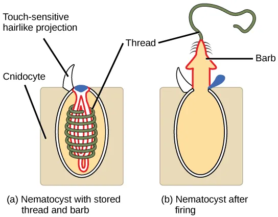
The defining cell type for the cnidarians is the cnidocyte, or stinging cell. These cells are located around the mouth and on the tentacles, and serve to capture prey or repel predators. Cnidocytes have large stinging organelles called nematocysts, which usually contain barbs at the base of a long coiled thread.

The illustration shows a nematocyst before, shown as a; and after, shown as b, firing. The nematocyst is a large, oval organelle inside a rectangular cnidocyte cell. The nematocyst is flush with the plasma membrane, and a touch-sensitive hairlike projection extends from the nematocyst to the cell's exterior. Inside the nematocyst, a thread is coiled around an inverted barb. Upon firing, a lid on the nematocyst opens. The barb pops out of the cell and the thread uncoils.
Figure 28.5 Cnidocytes. Animals from the phylum Cnidaria have stinging cells called cnidocytes. Cnidocytes contain large organelles called (a) nematocysts that store a coiled thread and barb, the nematocyst. When the hairlike cnidocil on the cell surface is touched, even lightly, (b) the thread, barb, and a toxin are fired from the organelle.

Link to Learning

View this video animation showing two anemones engaged in a battle.

Download Transcript

Morphology of Cnidarians

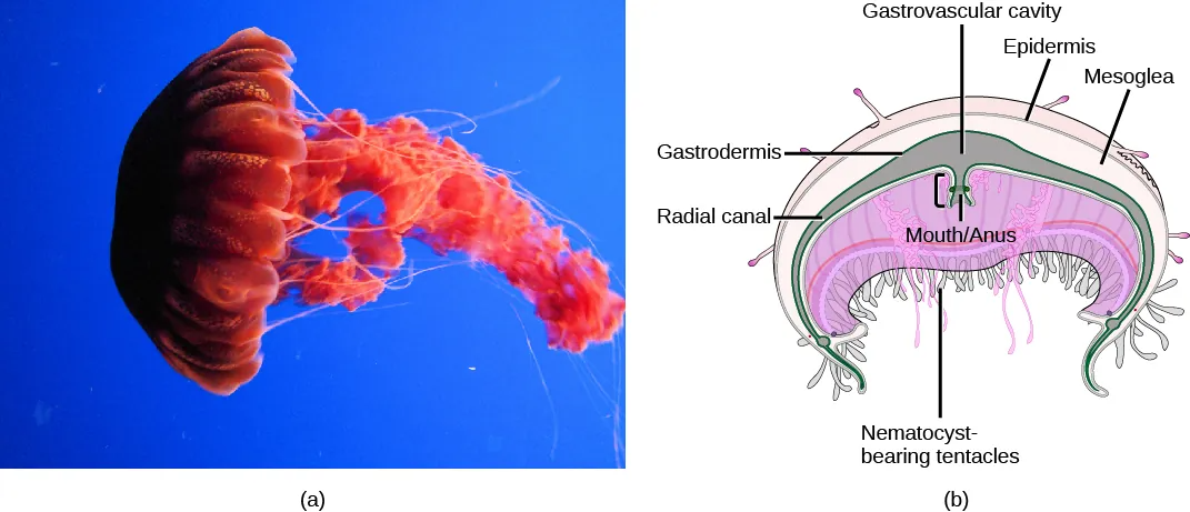
Two distinct body plans are found in Cnidarians: the polyp or tuliplike “stalk” form and the medusa or “bell” form. (Figure 28.6). An example of the polyp form is found in the genus Hydra, whereas the most typical form of medusa is found in the group called the “sea jellies” (jellyfish).  Polyp forms are sessile as adults, with a single opening (the mouth/anus) to the digestive cavity facing up with tentacles surrounding it. Medusa forms are motile, with the mouth and tentacles hanging down from an umbrella-shaped bell.

The illustration compares the medusa (a) and polyp (b) body plans. The medusa is dome-shaped, with tentacle-like appendages hanging down from the edges of the dome. The polyp looks like a tree, with a trunk at the bottom and branches at the top. Both the medusa and polyp have two tissue layers, with mesoglea in between. The mesoglea is thicker in the dome of the medusa than in the polyp. Both also have a central body cavity.
Figure 28.6 Cnidarian body forms. Cnidarians have two distinct body plans, the medusa (a) and the polyp (b). All cnidarians have two membrane layers, with a jelly-like mesoglea between them.

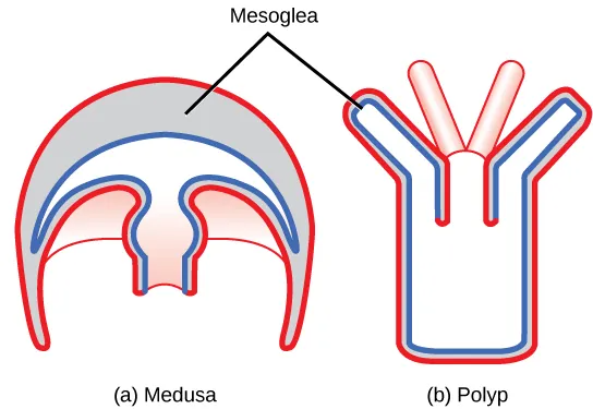
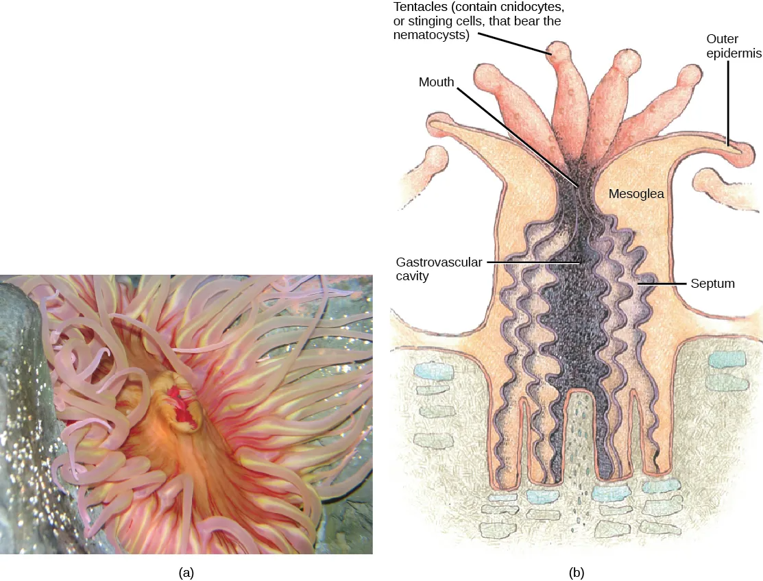
All cnidarians are diploblastic and thus have two “epithelial” layers in the body that are derived from the endoderm and ectoderm of the embryo. The outer layer (from ectoderm) is called the epidermis and lines the outside of the animal, whereas the inner layer (from endoderm) is called the gastrodermis and lines the digestive cavity. In the planula larva, a layer of ectoderm surrounds a solid mass of endoderm, but as the polyp develops, the digestive or gastrovascular cavity opens within the endoderm. A non-living, jelly-like mesoglea lies between these two epithelial layers. In terms of cellular complexity, cnidarians show the presence of differentiated cell types in each tissue layer, such as nerve cells, contractile epithelial cells, enzyme-secreting cells, and nutrient-absorbing cells, as well as the presence of intercellular connections. However, the development of organs or organ systems is not advanced in this phylum.

Physiological Processes of Cnidarians

The nervous system is rudimentary, with nerve cells organized in a network scattered across the body.

The gastrovascular cavity has only one opening that serves as both a mouth and an anus; this arrangement is called an incomplete digestive system. In the gastrovascular cavity, extracellular digestion occurs as food is taken into the gastrovascular cavity, enzymes are secreted into the cavity, and the cells lining the cavity absorb nutrients. However, some intracellular digestion also occurs. The gastrovascular cavity distributes nutrients throughout the body of the animal, with nutrients passing from the digestive cavity across the mesoglea to the epidermal cells. Thus, this cavity serves both digestive and circulatory functions.

Cnidarian cells exchange oxygen and carbon dioxide by diffusion between cells in the epidermis and water in the environment, and between cells in the gastrodermis and water in the gastrovascular cavity. The lack of a circulatory system to move dissolved gases limits the thickness of the body wall and necessitates a non-living mesoglea between the layers. In the cnidarians with a thicker mesoglea, a number of canals help to distribute both nutrients and gases. There is neither an excretory system nor organs, and nitrogenous wastes simply diffuse from the cells into the water outside the animal or into the gastrovascular cavity.

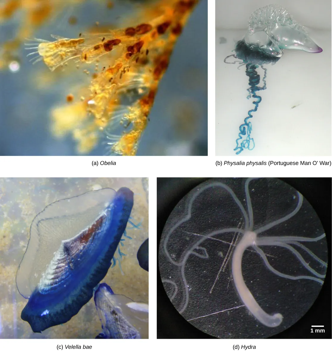
The phylum Cnidaria contains about 10,000 described species divided into two monophyletic clades: the Anthozoa and the Medusozoa. The Anthozoa include the corals, sea fans, sea whips, and the sea anemones. The Medusozoa include several classes of Cnidaria in two clades: The Hydrozoa include sessile forms, some medusoid forms, and swimming colonial forms like the Portuguese man-of-war. The other clade contains various types of jellies including both Scyphozoa (jellyfish) and Cubozoa (box jellies) The Anthozoa contain only sessile polyp forms, while the Medusozoa include species with both polyp and medusa forms in their life cycle.

Class Anthozoa:  Corals, sea fans, sea whips, sea anemones

Part a shows a photo of a sea anemone with a pink, oval body surrounded by thick, waving tentacles. Part b shows a cross-section of a sea anemone, which has a tube-shaped body with an opening called a gastrovascular cavity at its center. Ribbon-like septa divide this cavity into segments. A mesogleal layer separates the inner surface of the anemone from the outer surface. A mouth is located at the top of the gastrovascular cavity. Tentacles that contain stinging cnidocytes surround the mouth.
Figure 28.8 Sea anemone. The sea anemone is shown (a) photographed and (b) in a diagram illustrating its morphology. (credit a: modification of work by “Dancing With Ghosts”/Flickr; credit b: modification of work by NOAA)

Class Scyphozoa:  Jellyfish

Part a shows a photo of a bright red jellyfish with a dome-shaped body. Long tentacles drift from the bottom edge of the dome, and ribbon-like appendages trail from the middle of the body. Part b shows a cross-section of a jellyfish, which has nematocyst-bearing tentacles hanging from the bottom of the dome. Underneath the middle of the dome is an opening that serves as both a mouth and an anus. The opening leads to a gastrovascular cavity that is lined with a gastrodermis. The outer surface of the body is covered with an epidermis. Between the epidermis and gastrodermis is the mesoglea.
Figure 28.9 A sea jelly. A jelly is shown (a) photographed and (b) in a diagram illustrating its morphology. (credit a: modification of work by “Jimg944″/Flickr; credit b: modification of work by Mariana Ruiz Villareal)

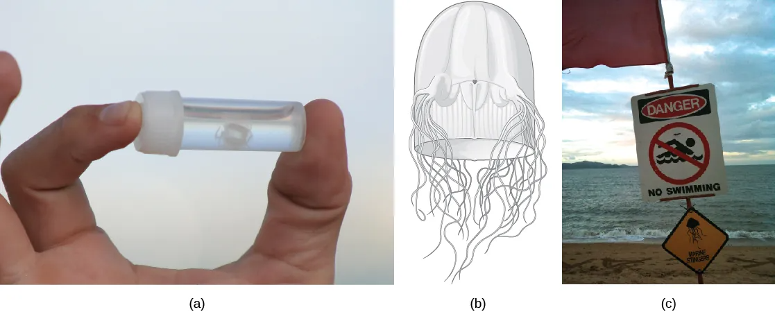
Class Cubozoa: Box Jellies

Photo A shows a person holding a small vial with a white jelly inside. The jelly is no bigger than a human fingernail. Illustration B shows a thimble-shaped jelly with two thick protrusions visible on either side. Tentacles radiate from the protrusions, and more tentacles radiate from the back. Photo C shows a sign posted on a beach beside the ocean that reads,danger, no swimming; with a picture of a jelly.
Figure 28.11 A cubozoan. The (a) tiny cubozoan jelly Malo kingi is thimble-shaped and, like all cubozoan jellies, (b) has four muscular pedalia to which the tentacles attach. M. kingi is one of two species of jellies known to cause Irukandji syndrome, a condition characterized by excruciating muscle pain, vomiting, increased heart rate, and psychological symptoms. Two people in Australia, where Irukandji jellies are most commonly found, are believed to have died from Irukandji stings. (c) A sign on a beach in northern Australia warns swimmers of the danger. (credit c: modification of work by Peter Shanks)

Class Hydrozoa:  Hydras, Siphophores (Portuguese Man o’ War)

Photo a shows Obelia, which has a body composed of branching polyps. Photo b shows a Portuguese Man O War, which has ribbon-like tentacles dangling from a clear, bulbous structure, resembling an inflated plastic bag. Photo c shows Velella bae, which resembles a flying saucer with a blue bottom and a clear, dome-shaped top. Photo d shows a hydra with long tentacles, extending from a tube-shaped body.
Figure 28.12 Hydrozoans. The polyp colony Obelia (a), siphonophore colonies Physalia (b) physalis, known as the Portuguese man o‘ war and Velella bae (c), and the solitary polyp Hydra (d) have different body shapes but all belong to the family Hydrozoa. (credit b: modification of work by NOAA; scale-bar data from Matt Russell)

License

Icon for the Creative Commons Attribution-ShareAlike 4.0 International License

General Biology Copyright © by Mary Ann Clark; Matthew Douglas; and Jung Choi is licensed under a Creative Commons Attribution-ShareAlike 4.0 International License, except where otherwise noted.

Share This Book