
21.2 Virus Infections and Hosts
Learning Outcomes
- List the steps of virus replication and explain what occurs at each step
- Describe the lytic and lysogenic cycles of virus replication
- Explain the transmission of plant and animal viruses
Viruses are obligate, intracellular parasites. A virus must first recognize and attach to a specific living cell prior to entering it. After penetration, the invading virus must copy its genome and manufacture its own proteins. Finally, the progeny virions must escape the host cell so that they can infect other cells. Viruses can infect only certain species of hosts and only certain cells within that host. Specific host cells that a virus must occupy and use to replicate are called permissive. In most cases, the molecular basis for this specificity is due to a particular surface molecule known as the viral receptor on the host cell surface. A specific viral receptor is required for the virus to attach. In addition, differences in metabolism and host-cell immune responses (based on differential gene expression) are a likely factor in determining which cells a virus may target for replication.
Steps of Virus Infections
A virus must use its host-cell processes to replicate. The viral replication cycle can produce dramatic biochemical and structural changes in the host cell, which may cause cell damage. These changes, called cytopathic effects, can change cell functions or even destroy the cell. Some infected cells, such as those infected by the common cold virus known as rhinovirus, die through lysis (bursting) or apoptosis (programmed cell death or “cell suicide”), releasing all progeny virions at once. The symptoms of viral diseases result both from such cell damage caused by the virus and from the immune response to the virus, which attempts to control and eliminate the virus from the body.
Many animal viruses, such as HIV (human immunodeficiency virus), leave the infected cells of the immune system by a process known as budding, where virions leave the cell individually. During the budding process, the cell does not undergo lysis and is not immediately killed. However, the damage to the cells that the virus infects may make it impossible for the cells to function normally, even though the cells remain alive for a period of time.
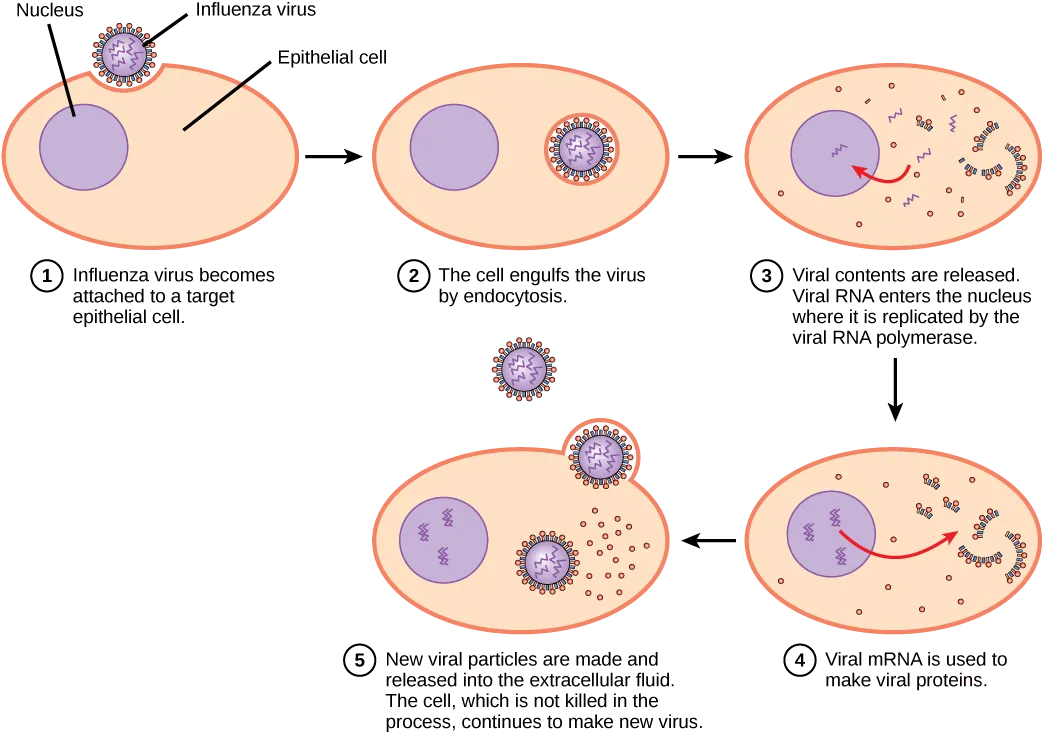
Most productive viral infections follow similar steps in the virus replication cycle (Figure 21.8):
- attachment
- penetration
- uncoating
- replication
- assembly
- release
Attachment
A virus attaches to a specific receptor site on the host cell membrane through attachment proteins in the capsid or via glycoproteins embedded in the viral envelope. The specificity of this interaction determines the host—and the cells within the host—that can be infected by a particular virus. This can be illustrated by thinking of several keys and several locks, where each key will fit only one specific lock.
Entry
Viruses may enter a host cell either with or without the viral capsid. The nucleic acid of bacteriophages enters the host cell “naked,” leaving the capsid outside the cell. Plant and animal viruses can enter through endocytosis (as you may recall, the cell membrane surrounds and engulfs the entire virus). Some enveloped viruses enter the cell when the viral envelope fuses directly with the cell membrane. Once inside the cell, the viral capsid degrades, and then the viral nucleic acid is released and becomes available for replication and transcription.
Replication and Assembly
The replication mechanism depends on the viral genome. DNA viruses usually use host-cell proteins and enzymes to replicate the viral DNA and to transcribe viral mRNA, which is then used to direct viral protein synthesis. RNA viruses usually use the RNA core as a template for synthesis of viral genomic RNA and mRNA. The viral mRNA directs the host cell to synthesize viral enzymes and capsid proteins, and assemble new virions.
Of course, there are exceptions to this pattern. If a host cell does not provide the enzymes necessary for viral replication, viral genes supply the information to direct synthesis of the missing proteins. Retroviruses, such as HIV (group VI of the Baltimore classification scheme), have an RNA genome that must be reverse transcribed into DNA, which then is incorporated into the host cell genome. To convert RNA into DNA, retroviruses must contain genes that encode the virus-specific enzyme reverse transcriptase that transcribes an RNA template to DNA. Reverse transcription never occurs in uninfected host cells—the enzyme reverse transcriptase is only derived from the expression of viral genes within the infected host cells. The fact that HIV produces some of its own enzymes not found in the host has allowed researchers to develop drugs that inhibit these enzymes without affecting the host’s metabolism.
This approach has led to the development of a variety of drugs used to treat HIV and has been effective at reducing the number of infectious virions (copies of viral RNA) in the blood to non-detectable levels in many HIV-infected individuals.
Egress
The last stage of viral replication is the release of the new virions produced in the host organism, where they are able to infect adjacent cells and repeat the replication cycle. As you’ve learned, some viruses are released when the host cell dies, and other viruses can leave infected cells by budding through the membrane without directly killing the cell.
Link to Learning
Virus infection
This video explains how influenza attacks the body.
Link to Learning
Virus transmission and replication
Watch a video on viruses, identifying structures, modes of transmission, replication, and more.
Different Hosts and Their Viruses
As you’ve learned, viruses often infect very specific hosts, as well as specific cells within the host. This feature of a virus makes it specific to one or a few species of life on Earth. On the other hand, so many different types of viruses exist on Earth that nearly every living organism has its own set of viruses trying to infect its cells. Even prokaryotes, the smallest and simplest of cells, may be attacked by specific types of viruses. In the following section, we will look at some of the features of viral infection of prokaryotic cells. As we have learned, viruses that infect bacteria are called bacteriophages (Figure 21.9). Archaea have their own similar viruses.
Bacteriophages
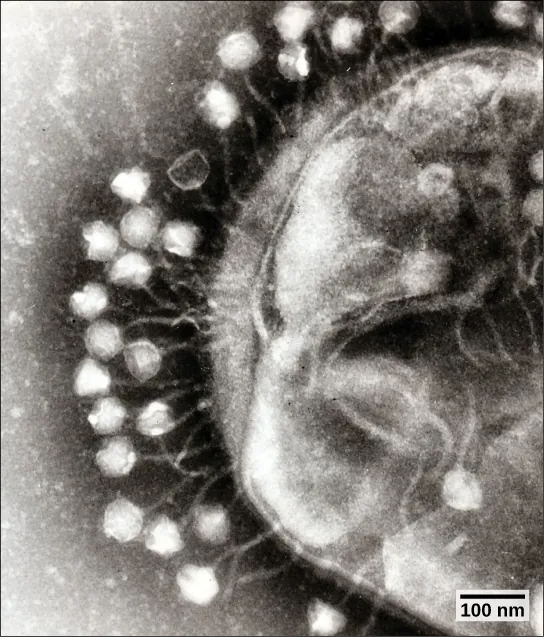
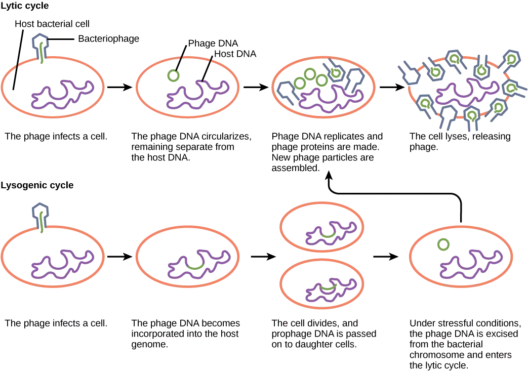
Most bacteriophages are dsDNA viruses, which use host enzymes for DNA replication and RNA transcription. Phage particles must bind to specific surface receptors and actively insert the genome into the host cell. (The complex tail structures seen in many bacteriophages are actively involved in getting the viral genome across the prokaryotic cell wall.) When infection of a cell by a bacteriophage results in the production of new virions, the infection is said to be productive. If the virions are released by bursting the cell, the virus replicates by means of a lytic cycle (Figure 21.10). An example of a lytic bacteriophage is T4, which infects Escherichia coli found in the human intestinal tract. Sometimes, however, a virus can remain within the cell without being released. For example, when a temperate bacteriophage infects a bacterial cell, it replicates by means of a lysogenic cycle (Figure 21.10), and the viral genome is incorporated into the genome of the host cell. When the phage DNA is incorporated into the host-cell genome, it is called a prophage. An example of a lysogenic bacteriophage is the λ (lambda) virus, which also infects the E. coli bacterium. Viruses that infect plant or animal cells may sometimes undergo infections where they are not producing virions for long periods. An example is the animal herpesviruses, including herpes simplex viruses, the cause of oral and genital herpes in humans. In a process called latency, these viruses can exist in nervous tissue for long periods of time without producing new virions, only to leave latency periodically and cause lesions in the skin where the virus replicates. Even though there are similarities between lysogeny and latency, the term lysogenic cycle is usually reserved to describe bacteriophages.
Plant Viruses
Most plant viruses, like the tobacco mosaic virus, have single-stranded (+) RNA genomes. However, there are also plant viruses in most other virus categories. Unlike bacteriophages, plant viruses do not have active mechanisms for delivering the viral genome across the protective cell wall. For a plant virus to enter a new host plant, some type of mechanical damage must occur. This damage is often caused by weather, insects, animals, fire, or human activities like farming or landscaping. Movement from cell to cell within a plant can be facilitated by viral modification of plasmodesmata (cytoplasmic threads that pass from one plant cell to the next). Additionally, plant offspring may inherit viral diseases from parent plants. Plant viruses can be transmitted by a variety of vectors, through contact with an infected plant’s sap, by living organisms such as insects and nematodes, and through pollen. The transfer of a virus from one plant to another is known as horizontal transmission, whereas the inheritance of a virus from a parent is called vertical transmission.
Plant viruses can seriously disrupt crop growth and development, significantly affecting our food supply. They are responsible for poor crop quality and quantity globally, and can bring about huge economic losses annually.
Animal Viruses
Animal viruses, unlike the viruses of plants and bacteria, do not have to penetrate a cell wall to gain access to the host cell. The virus may even induce the host cell to cooperate in the infection process. Non-enveloped or “naked” animal viruses may enter cells in two different ways. As a protein in the viral capsid binds to its receptor on the host cell, the virus may be taken inside the cell via a vesicle during the normal cell process of receptor-mediated endocytosis. An alternative method of cell penetration used by non-enveloped viruses is for capsid proteins to undergo shape changes after binding to the receptor, creating channels in the host cell membrane. The viral genome is then “injected” into the host cell through these channels in a manner analogous to that used by many bacteriophages.
Enveloped viruses also have two ways of entering cells after binding to their receptors: receptor-mediated endocytosis, or fusion. Many enveloped viruses enter the cell by receptor-mediated endocytosis in a fashion similar to that seen in some non-enveloped viruses. On the other hand, fusion only occurs with enveloped virions. These viruses, which include HIV among others, use special fusion proteins in their envelopes to cause the envelope to fuse with the plasma membrane of the cell, thus releasing the genome and capsid of the virus into the cell cytoplasm.
After making their proteins and copying their genomes, animal viruses complete the assembly of new virions and exit the cell. As we have already discussed using the example the influenza virus, enveloped animal viruses may bud from the cell membrane as they assemble themselves, taking a piece of the cell’s plasma membrane in the process. On the other hand, non-enveloped viral progeny, such as rhinoviruses, accumulate in infected cells until there is a signal for lysis or apoptosis, and all virions are released together.
Link to Learning
Lytic and Lysogenic Cycle
Watch a video on the lytic and lysogenic life cycles.
cell type that is able to support productive replication of a virus
causing cell damage
bursting of a cell
method of exit from the cell used in certain animal viruses, where virions leave the cell individually by capturing a piece of the host plasma membrane
virus that infects bacteria
type of virus replication in which virions are released through lysis, or bursting, of the cell
type of virus replication in which the viral genome is incorporated into the genome of the host cell
transmission of a disease between unrelated individuals
transmission of disease from parent to offspring
variation of endocytosis that involves the use of specific binding proteins in the plasma membrane for specific molecules or particles, and clathrin-coated pits that become clathrin-coated vesicles
method of entry by some enveloped viruses, where the viral envelope fuses with the plasma membrane of the host cell
