32.1 Reproductive Development and Structure
Learning Outcomes
- Describe the two stages of a plant’s life cycle
- Compare and contrast male and female gametophytes and explain how they form in angiosperms
- Describe the reproductive structures of a plant
- Describe the components of a complete flower
- Describe the development of microsporangium and megasporangium in gymnosperms
Sexual reproduction takes place with slight variations in different groups of plants. Plants have two distinct stages in their lifecycle: the gametophyte stage and the sporophyte stage. The haploid gametophyte produces the male and female gametes by mitosis in distinct multicellular structures. Fusion of the male and females gametes forms the diploid zygote, which develops into the sporophyte. After reaching maturity, the diploid sporophyte produces spores by meiosis, which in turn divide by mitosis to produce the haploid gametophyte. The new gametophyte produces gametes, and the cycle continues. This is the alternation of generations, and is typical of plant reproduction (Figure 32.2).
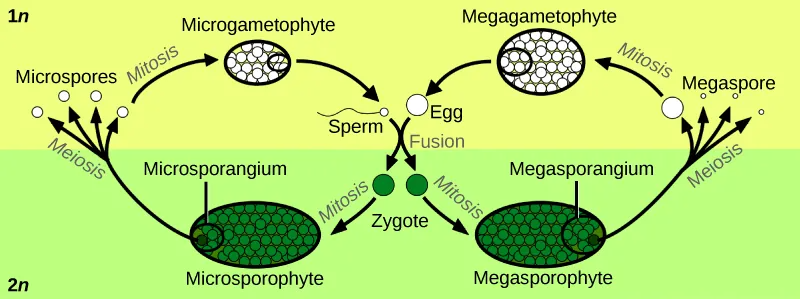
The life cycle of higher plants is dominated by the sporophyte stage, with the gametophyte borne on the sporophyte. In ferns, the gametophyte is free-living and very distinct in structure from the diploid sporophyte. In bryophytes, such as mosses, the haploid gametophyte is more developed than the sporophyte.
During the vegetative phase of growth, plants increase in size and produce a shoot system and a root system. As they enter the reproductive phase, some of the branches start to bear flowers. The flower is borne on a stalk known as a receptacle. Flower shape, color, and size are unique to each species, and are often used by taxonomists to classify plants.
Sexual Reproduction in Angiosperms
The lifecycle of angiosperms follows the alternation of generations explained previously. The haploid gametophyte alternates with the diploid sporophyte during the sexual reproduction process of angiosperms. Flowers contain the plant’s reproductive structures.
Flower Structure
A typical flower has four main parts, or whorls, known as the calyx, corolla, androecium, and gynoecium (Figure 32.3). The outermost whorl of the flower has green, leafy structures known as sepals. The sepals, collectively called the calyx, help to protect the unopened bud. The second whorl is comprised of petals—usually, brightly colored—collectively called the corolla. The number of sepals and petals varies depending on whether the plant is a monocot or dicot. In monocots, petals usually number three or multiples of three; in dicots, the number of petals is four or five, or multiples of four and five. Together, the calyx and corolla are known as the perianth. The third whorl contains the male reproductive structures and is known as the androecium. The androecium has stamens with anthers that contain the microsporangia. The innermost group of structures in the flower is the gynoecium, or the female reproductive component(s). The carpel is the individual unit of the gynoecium and has a stigma, style, and ovary. A flower may have one or multiple carpels.
Visual Connection
Flower Anatomy
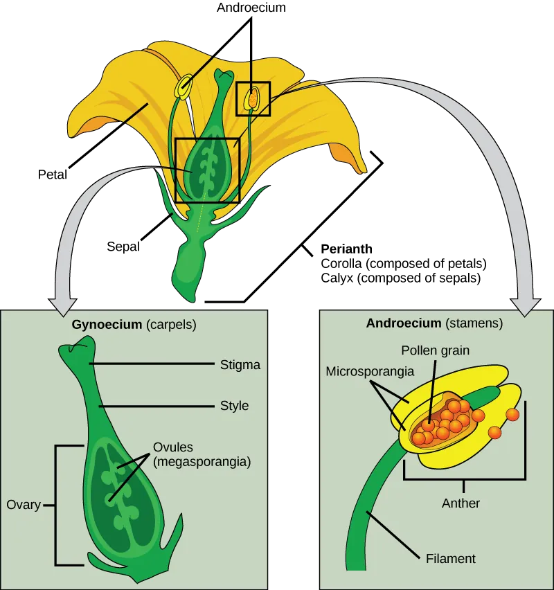
If all four whorls (the calyx, corolla, androecium, and gynoecium) are present, the flower is described as complete. If any of the four parts is missing, the flower is known as incomplete. Flowers that contain both an androecium and a gynoecium are called perfect, androgynous or hermaphrodites. There are two types of incomplete flowers: staminate flowers contain only stamen, and carpellate flowers have only carpels (Figure 32.4).
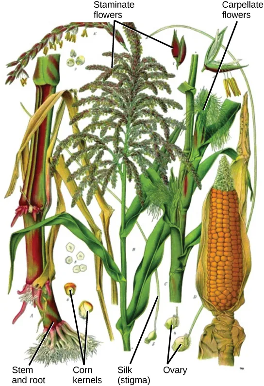
If both male and female flowers are borne on the same plant, the species is called monoecious (meaning “one home”). Species with male and female flowers borne on separate plants are termed dioecious, or “two homes.”
Male Gametophyte (The Pollen Grain)
The male gametophyte develops and reaches maturity in an immature anther. In a plant’s male reproductive organs, development of pollen takes place in a structure known as the microsporangium (Figure 32.6). The microsporangia are pollen sacs in which the microspores develop into pollen grains. These are found in the anther, which is at the end of the stamen—the long filament that supports the anther.
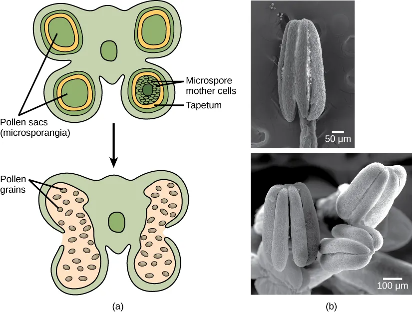
Within the microsporangium, the microspore mother cell divides by meiosis to give rise to four microspores, each of which will ultimately form a pollen grain (Figure 32.7). An inner layer of cells, known as the tapetum, provides nutrition to the developing microspores and contributes key components to the pollen wall. Mature pollen grains contain two cells: a generative cell and a pollen tube cell. The generative cell is contained within the larger pollen tube cell. Upon germination, the tube cell forms the pollen tube through which the generative cell migrates to enter the ovary. During its transit inside the pollen tube, the generative cell divides to form two male gametes (sperm cells). Upon maturity, the microsporangia burst, releasing the pollen grains from the anther.
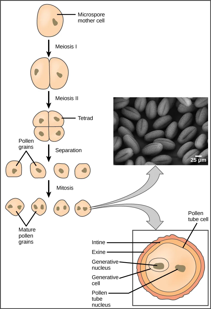
Each pollen grain has a covering that contains sporopollenin, a complex waterproofing substance that allows the pollen to survive under unfavorable conditions and to be carried by wind, water, or biological agents without undergoing damage.
Female Gametophyte (The Embryo Sac)
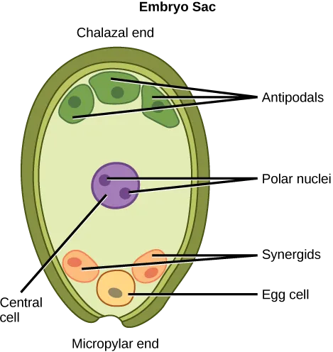
While the details may vary between species, the overall development of the female gametophyte has two distinct phases. First, in the process of megasporogenesis, a single cell in the diploid megasporangium undergoes meiosis to produce four megaspores, only one of which survives. During the second phase, megagametogenesis, the surviving haploid megaspore undergoes mitosis, but without cytokinesis, to produce an eight-nucleate, seven-cell female gametophyte, also known as the embryo sac. Two of the nuclei—the polar nuclei—move to the equator and fuse, forming a single, diploid central cell. This central cell later fuses with a sperm to form the triploid endosperm. Three nuclei position themselves on the end of the embryo sac opposite the micropyle and develop into the antipodal cells, which later degenerate. The nucleus closest to the micropyle becomes the female gamete, or egg cell, and the two adjacent nuclei develop into synergid cells (Figure 32.8). The synergids help guide the pollen tube for successful fertilization, after which they disintegrate. Once fertilization is complete, the resulting diploid zygote develops into the embryo, and the fertilized ovule forms the other tissues of the seed.
A double-layered integument protects the megasporangium and, later, the embryo sac. The integument will develop into the seed coat after fertilization and protect the entire seed. The ovule wall will become part of the fruit. The integuments, while protecting the megasporangium, do not enclose it completely, but leave an opening called the micropyle. The micropyle allows the pollen tube to enter the female gametophyte for fertilization.
Visual Connection
Embryo Sac
Sexual Reproduction in Gymnosperms
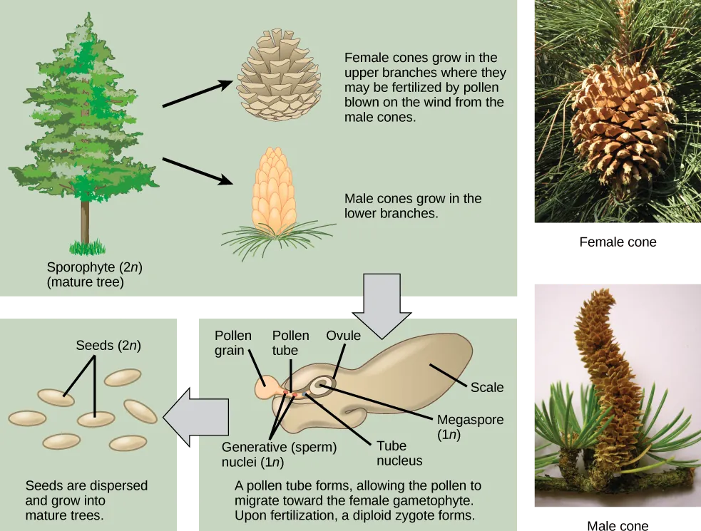
As with angiosperms, the lifecycle of a gymnosperm is also characterized by alternation of generations. In conifers such as pines, the green leafy part of the plant is the sporophyte, and the cones contain the male and female gametophytes (Figure 32.9). The female cones are larger than the male cones and are positioned towards the top of the tree; the small, male cones are located in the lower region of the tree. Because the pollen is shed and blown by the wind, this arrangement makes it difficult for a gymnosperm to self-pollinate.
Male Gametophyte
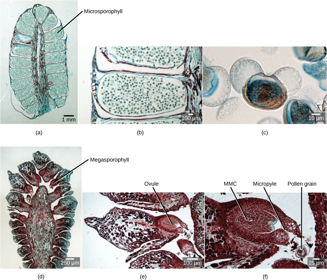
A male cone has a central axis on which bracts, a type of modified leaf, are attached. The bracts are known as microsporophylls (Figure 32.10) and are the sites where microspores will develop. The microspores develop inside the microsporangium. Within the microsporangium, cells known as microsporocytes divide by meiosis to produce four haploid microspores. Further mitosis of the microspore produces two nuclei: the generative nucleus, and the tube nucleus. Upon maturity, the male gametophyte (pollen) is released from the male cones and is carried by the wind to land on the female cone.
Female Gametophyte
The female cone also has a central axis on which bracts known as megasporophylls (Figure 32.10) are present. In the female cone, megaspore mother cells are present in the megasporangium. The megaspore mother cell divides by meiosis to produce four haploid megaspores. One of the megaspores divides to form the multicellular female gametophyte, while the others divide to form the rest of the structure. The female gametophyte is contained within a structure called the archegonium.
Reproductive Process
Upon landing on the female cone, the tube cell of the pollen forms the pollen tube, through which the generative cell migrates towards the female gametophyte through the micropyle. It takes approximately one year for the pollen tube to grow and migrate towards the female gametophyte. The male gametophyte containing the generative cell splits into two sperm nuclei, one of which fuses with the egg, while the other degenerates. After fertilization of the egg, the diploid zygote is formed, which divides by mitosis to form the embryo. The scales of the cones are closed during development of the seed. The seed is covered by a seed coat. Seed development takes another one to two years. Once the seed is ready to be dispersed, the bracts of the female cones open to allow the dispersal of seed.

