28.4 Phylum Mollusca and Annelida
Learning Outcomes
- Describe the unique anatomical and morphological features of molluscs and annelids
The annelids and the mollusks are the most familiar of the lophotrochozoan protostomes. They are also more “typical” lophotrochozoans, since both groups include aquatic species with trochophore larvae, which unite both taxa in common ancestry. These phyla show how a flexible body plan can lead to biological success in terms of abundance and species diversity. The phylum Mollusca has the second greatest number of species of all animal phyla with nearly 100,000 described extant species, and about 80,000 described extinct species. In fact, it is estimated that about 25 percent of all known marine species are mollusks! The annelids and mollusca are both bilaterally symmetrical, cephalized, triploblastic, schizocoelous eucoeolomates. They include animals you are likely to see in your backyard or on your dinner plate!
Phylum Mollusca
The name “Mollusca” means “soft” body, since the earliest descriptions of molluscs came from observations of “squishy,” unshelled cuttlefish. Molluscs are predominantly a marine group of animals; however, they are also known to inhabit freshwater as well as terrestrial habitats. This enormous phylum includes chitons, tusk shells, snails, slugs, nudibranchs, sea butterflies, clams, mussels, oysters, squids, octopuses, and nautiluses. Molluscs display a wide range of morphologies in each class and subclass, but share a few key characteristics (Figure 28.21). The chief locomotor structure is usually a muscular foot. Most internal organs are contained in a region called the visceral mass. Overlying the visceral mass is a fold of tissue called the mantle; within the cavity formed by the mantle are respiratory structures called gills, that typically fold over the visceral mass. The mouths of most mollusks, except bivalves (e.g., clams) contain a specialized feeding organ called a radula, an abrasive tonguelike structure. Finally, the mantle secretes a calcium-carbonate-hardened shell in most molluscs, although this is greatly reduced in the class Cephalopoda, which contains the octopuses and squids.
Visual Connection
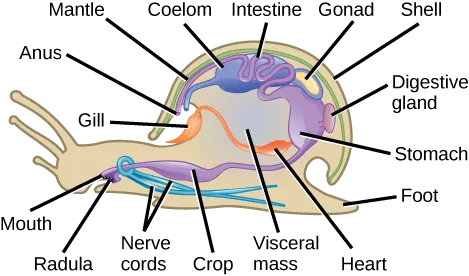
Classification of Phylum Mollusca
Phylum Mollusca comprises a very diverse group of organisms that exhibits a dramatic variety of forms, ranging from chitons to snails to squids, the latter of which typically show a high degree of intelligence. This variability is a consequence of modification of the basic body regions, especially the foot and mantle. The following will outline some of the more commonly known classes of mollusks and their distinguishing characteristics.
Class Bivalvia
Class Bivalvia (“two-valves”) includes clams, oysters, mussels, scallops, geoducks, and shipworms. Some bivalves are almost microscopic, while others, in the genus Tridacna, may be one meter in length and weigh 225 kilograms. Members of this class are found in marine as well as freshwater habitats. As the name suggests, bivalves are enclosed in two-part valves or shells (Figure 28.23a) fused on the dorsal side by hinge ligaments as well as shell teeth on the ventral side that keep the two halves aligned. The two shells are joined at the oldest part of the shell called the umbo. Anterior and posterior adductor and abductor muscles close and open the shell respectively.
The overall body of the bivalve is laterally flattened; the foot is wedge-shaped; and the head region is poorly developed (with no obvious mouth). Bivalves are filter-feeders, and a radula is absent in this class of mollusks. The mantle cavity is fused along the edges except for openings for the foot and for the intake and expulsion of water, which is circulated through the mantle cavity by the actions of the incurrent and excurrent siphons. During water intake by the incurrent siphon, food particles are captured by the paired posterior gills (ctenidia) and then carried by the movement of cilia forward to the mouth. Excretion and osmoregulation are performed by a pair of nephridia. Eyespots and other sensory structures are located along the edge of the mantle in some species. The “eyes” are especially conspicuous in scallops (Figure 28.23b). Three pairs of connected ganglia regulate activity of different body structures.

One of the functions of the mantle is to secrete the shell. Some bivalves, like oysters and mussels, possess the unique ability to secrete and deposit a calcareous nacre or “mother of pearl” around foreign particles that may enter the mantle cavity. This property has been commercially exploited to produce pearls.
Link to Learning
Did you know the scallops can swim? Watch this video of scallops “swimming”!
Class Gastropoda
More than half of molluscan species are in the class Gastropoda (“stomach foot”), which includes well-known mollusks like snails, slugs, conchs, cowries, limpets, and whelks. Aquatic gastropods include both marine and freshwater species, and all terrestrial mollusks are gastropods. Gastropoda includes shell-bearing species as well as species without shells. Gastropod bodies are asymmetrical and usually present a coiled shell (Figure 28.24a).
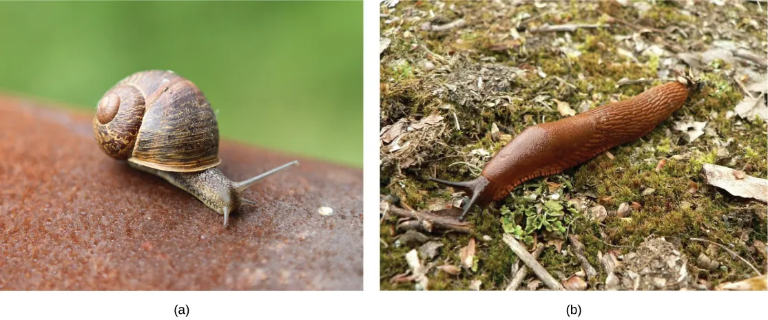
Gastropods have a foot that is modified for crawling. Most gastropods have a well-defined head with tentacles and eyes. A complex radula is used to scrape up food particles. In aquatic gastropods, the mantle cavity encloses the gills (ctenidia), but in land gastropods, the mantle itself is the major respiratory structure, acting as a kind of lung. Nephridia (“kidneys”) are also found in the mantle cavity.
Class Cephalopoda
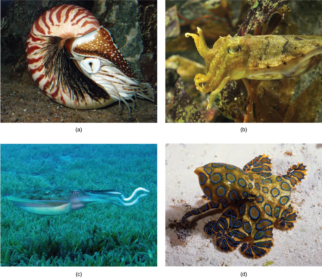
Class Cephalopoda (“head foot” animals), includes octopuses, squids, cuttlefish, and nautiluses. Cephalopods include both animals with shells as well as animals in which the shell is reduced or absent. In the shell-bearing Nautilus, the spiral shell is multi-chambered. These chambers are filled with gas or water to regulate buoyancy. The shell structure in squids and cuttlefish is reduced and is present internally in the form of a squid pen and cuttlefish bone, respectively. Cuttle bone is sold in pet stores to help smooth the beaks of birds and also to provide birds such as egg-laying chickens and quail with an inexpensive natural source of calcium carbonate. Examples of cephalopods are shown in Figure 28.27.
Cephalopods can display vivid and rapidly changing coloration, almost like flashing neon signs. Typically these flashing displays are seen in squids and octopuses, where they may be used for camouflage and possibly as signals for mating displays.
All animals in this class are carnivorous predators and have beak-like jaws in addition to the radula. Cephalopods include the most intelligent of the mollusks, and have a well-developed nervous system along with image-forming eyes. Unlike other mollusks, they have a closed circulatory system, in which the blood is entirely contained in vessels rather than in a hemocoel.
The foot is lobed and subdivided into arms and tentacles. Suckers with chitinized rings are present on the arms and tentacles of octopuses and squid. Siphons are well developed and the expulsion of water is used as their primary mode of locomotion, which resembles jet propulsion. Gills (ctenidia) are attached to the wall of the mantle cavity and are serviced by large blood vessels, each with its own heart. A pair of nephridia is present within the mantle cavity for water balance and excretion of nitrogenous wastes. Cephalopods such as squids and octopuses also produce sepia or a dark ink, which contains melanin. The ink gland is located between the gills and can be released into the excurrent water stream. Ink clouds can be used either as a “smoke screen” to hide the animal from predators during a quick attempt at escape, or to create a fake image to distract predators.
Cephalopods are dioecious. Members of a species mate, and the female then lays the eggs in a secluded and protected niche. Females of some species care for the eggs for an extended period of time and may end up dying during that time period. While most other aquatic mollusks produce trochophore larvae, cephalopod eggs develop directly into a juvenile without an intervening larval stage.
Phylum Annelida
Phylum Annelida comprises the true, segmented worms. These animals are found in marine, terrestrial, and freshwater habitats, but the presence of water or humidity is a critical factor for their survival in terrestrial habitats. The annelids are often called “segmented worms” due to their key characteristic of metamerism, or true segmentation. Approximately 16,500 species have been described in phylum Annelida, which includes polychaete worms (marine annelids with multiple appendages), and oligochaetes (earthworms and leeches). Some animals in this phylum show parasitic and commensal symbioses with other species in their habitat.
Morphology
Annelids display bilateral symmetry and are worm-like in overall morphology. The name of the phylum is derived from the Latin word annullus, which means a small ring, an apt description of the ring-like segmentation of the body. Annelids have a body plan with metameric segmentation, in which several internal and external morphological features are repeated in each body segment. Metamerism allows animals to become bigger by adding “compartments,” while making their movement more efficient.
Anatomy
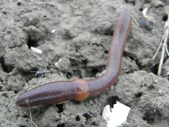
The epidermis is protected by a collagenous, external cuticle, which is much thinner than the cuticle found in the ecdysozoans and does not require periodic shedding for growth. Circular as well as longitudinal muscles are located interior to the epidermis. Chitinous bristles called setae (or chaetae) are anchored in the epidermis, each with its own muscle. In the polychaetes, the setae are borne on paired appendages called parapodia.
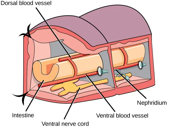
Most annelids have a well-developed and complete digestive system. Feeding mechanisms vary widely across the phylum. Some polychaetes are filter-feeders that use feather-like appendages to collect small organisms. Others have tentacles, jaws, or an eversible pharynx to capture prey. Earthworms collect small organisms from soil as they burrow through it, and most leeches are blood-feeders armed with teeth or a muscular proboscis. In earthworms, the digestive tract includes a mouth, muscular pharynx, esophagus, crop, and muscular gizzard. The gizzard leads to the intestine, which ends in an anal opening in the terminal segment. A cross-sectional view of a body segment of an earthworm is shown in Figure 28.30; each segment is limited by a membranous septum that divides the coelomic cavity into a series of compartments.
Most annelids possess a closed circulatory system of dorsal and ventral blood vessels that run parallel to the alimentary canal as well as capillaries that service individual tissues. Annelids lack a well-developed respiratory system, and gas exchange occurs across the moist body surface. In the polychaetes, the parapodia are highly vascular and serve as respiratory structures. Excretion is facilitated by a pair of metanephridia (a type of primitive “kidney” that consists of a convoluted tubule and an open, ciliated funnel) that is present in every segment toward the ventral side. Annelids show well-developed nervous systems with a ring of fused ganglia present around the pharynx. The nerve cord is ventral in position and bears enlarged nodes or ganglia in each segment.
Annelids may be either monoecious with permanent gonads (as in earthworms and leeches) or dioecious with temporary or seasonal gonads (as in polychaetes). However, cross-fertilization is preferred even in hermaphroditic animals. Earthworms may show simultaneous mutual fertilization when they are aligned for copulation. Some leeches change their sex over their reproductive lifetimes. In most polychaetes, fertilization is external and development includes a trochophore larva, which then metamorphoizes to the adult form. In oligochaetes, fertilization is typically internal and the fertilized eggs develop in a cocoon produced by the clitellum; development is direct. Polychaetes are excellent regenerators and some even reproduce asexually by budding or fragmentation.
Link to Learning
This combination video and animation provides a close-up look at annelid anatomy.

Link to Learning
For a nice summary of invertebrates, watch the Crash Course Biology video “Simple Animals: Sponges, Jellies & Octopuses.”

