28.3 Phylum Platyhelminthes (in superphylum Lophotrochozoa)
Learning Outcomes
- Describe the unique anatomical and morphological features of flatworms
Animals belonging to superphylum Lophotrochozoa are triploblastic (have three germ layers) and unlike the cnidarians, they possess an embryonic mesoderm sandwiched between the ectoderm and endoderm. These phyla are also bilaterally symmetrical, meaning that a longitudinal section will divide them into right and left sides that are superficially symmetrical. In these phyla, we also see the beginning of cephalization, the evolution of a concentration of nervous tissues and sensory organs in the head of the organism—exactly where a mobile bilaterally symmetrical organism first encounters its environment.
Lophotrochozoa are also protostomes, in which the blastopore, or the point of invagination of the ectoderm (outer germ layer), becomes the mouth opening into the alimentary canal. This developmental pattern is called protostomy or “first mouth.” Protostomes include acoelomate, pseudocoelomate, and eucoelomate phyla. The coelom is a cavity that separates the ectoderm from the endoderm. In acoelomates, a solid mass of mesoderm is sandwiched between the ectoderm and endoderm and does not form a cavity or “coelom,” leaving little room for organ development; in pseudocoelomates, there is a cavity or pseudocoelom that replaces the blastocoel (the cavity within the blastula), but it is only lined by mesoderm on the outside of the cavity, leaving the gut tube and organs unlined; in eucoelomates, the cavity that obliterates the blastocoel as the coelom develops is lined both on the outside of the cavity (parietal layer) and also on the inside of the cavity, around the gut tube and the internal organs (visceral layer).
Phylum Platyhelminthes
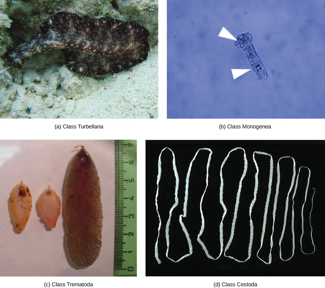
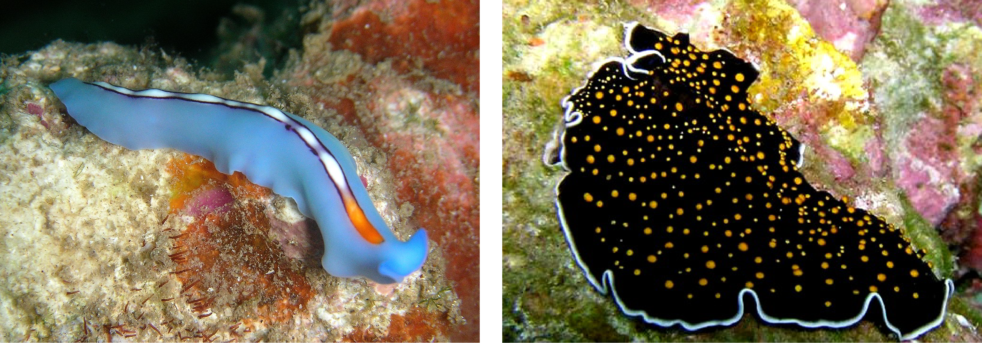
The flatworms are acoelomate organisms that include many free-living and parasitic forms (trematodes and cestodes/tapeworms).
Physiological Processes of Flatworms
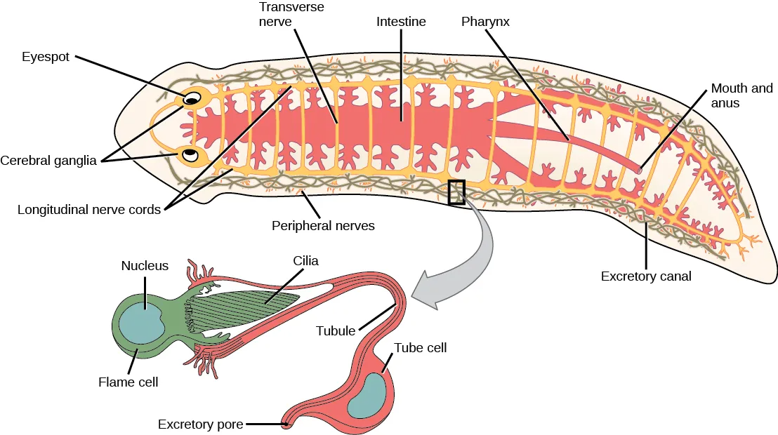
The free-living species of flatworms are predators or scavengers. Parasitic forms feed by absorbing nutrients provided by their hosts. Most flatworms, such as the planarian shown in Figure 28.14, have a branching gastrovascular cavity rather than a complete digestive system. In such animals, the “mouth” is also used to expel waste materials from the digestive system, and thus also serves as an anus. Digestion is primarily extracellular, with digested materials taken into the cells of the gut lining by phagocytosis. One parasitic group, the tapeworms (cestodes), lacks a digestive system altogether, and absorb digested food from the host.
Flatworms have an excretory system with a network of tubules attached to flame cells, whose cilia beat to direct waste fluids concentrated in the tubules out of the body through excretory pores. . The nervous system consists of a pair of lateral nerve cords running the length of the body with transverse connections between them. Two large cerebral ganglia—concentrations of nerve cell bodies at the anterior end of the worm—are associated with photosensory and chemosensory cells. There is neither a circulatory nor a respiratory system, with gas and nutrient exchange dependent on diffusion and cell-to-cell junctions. This necessarily limits the thickness of the body in these organisms, constraining them to be “flat” worms. Most flatworm species are monoecious (both male and female reproductive organs are found in the same individual), and fertilization is typically internal. Asexual reproduction by fission is common in some groups.